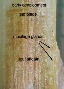
The presence and visibility of mucilage glands can be diagnostic for Typha species. Mucilage glands become more discernable as the plants age—going from clear to brown to black. The glands are seen on the inside of the leaf sheath and are often diagnostic at the sheath-leaf blade transition. They are roughly rectangular cells arranged in longitudinal columns which are more visible along the edge of the sheath and less-often visible along the center of the sheath or on the leaf blade. Staining the tissue with safranin will make the glands more visible.
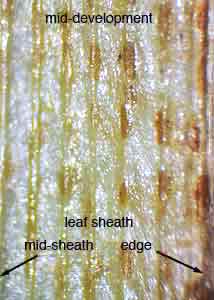
Typha angustifolia (narrowleaf cattail). This introduced species, has mucilage glands on the leaf sheath but they are absent from the central part of the sheath and from the leaf blade. During mid to late development, they become visible to the unaided eye on the sides and lower central part of the sheath. Reading from left to right; the first two pictures below show the adaxial surface of the leaf sheath on T. angustifolia
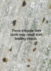
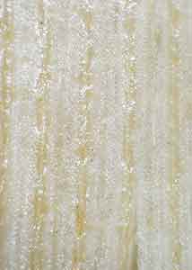
The next two pictures are from Typha latifolia (broadleaf cattail). This native species, has mucilage glands that are clear (last photo) and not visible to the unaided eye at any stage of development. It is easy to be misled by a type of insect damage that often occurs in the same region as the mucilage glands. These irregular brown/black necrotic spots are thought to be the work of feeding insects. Leaf mucilage glands are usually colorless and difficult to see in fresh leaves of both species early in the season and in Typha latifolia at all stages. They are brown and clearly evident to the unaided eye in mid- to late-season fresh or dried T. angustifolia and T. domingensis and are easily stained (with, e.g., safranin).